Blue/Green LED Technology
Safe Detection of all Red and Green DNA Dyes
The Blue/Green Revolution
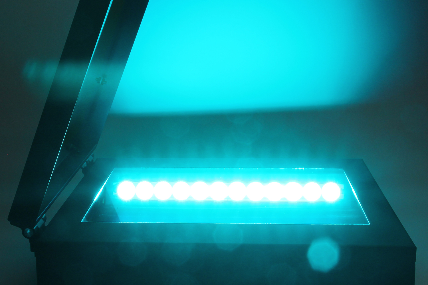
The Blue/Green LED technology is a safe and revolutionary method for the detection of stained nucleic acids. The technology uses a combination of wavelengths in a light spectrum ranging from 470 nm to 520 nm. This light range lies in the visible light spectrum and is not harmful for the user or for the DNA sample. As the fundamental part of our basic and advanced gel documentation systems, Blue/Green LEDs efficiently excite all common red and green DNA dyes (such as ethidium bromide or MIDORIGreen) with a very high intensity. On this page, we would like to demonstrate the biggest downsides of commonly used UV-light for DNA visualization, explain the advantages and revolutionary technology behind Blue/Green LED light and introduce our impressive Blue/Green LED Gel Documentation systems.
Danger of UV-light
UV-light, together with ethidium bromide staining, has been commonly used to detect nucleic acids in molecular biology laboratories for 50 years and is still used to this day. However, DNA absorbs light in the UV-spectrum causing strong damage to DNA and leading to modifications and degradation, not only for the DNA sample but also for the user. We are convinced, it is time to move on from this damaging and dangerous light source, as DNA visualization with UV-light does not represent the state-of-the-art method anymore.
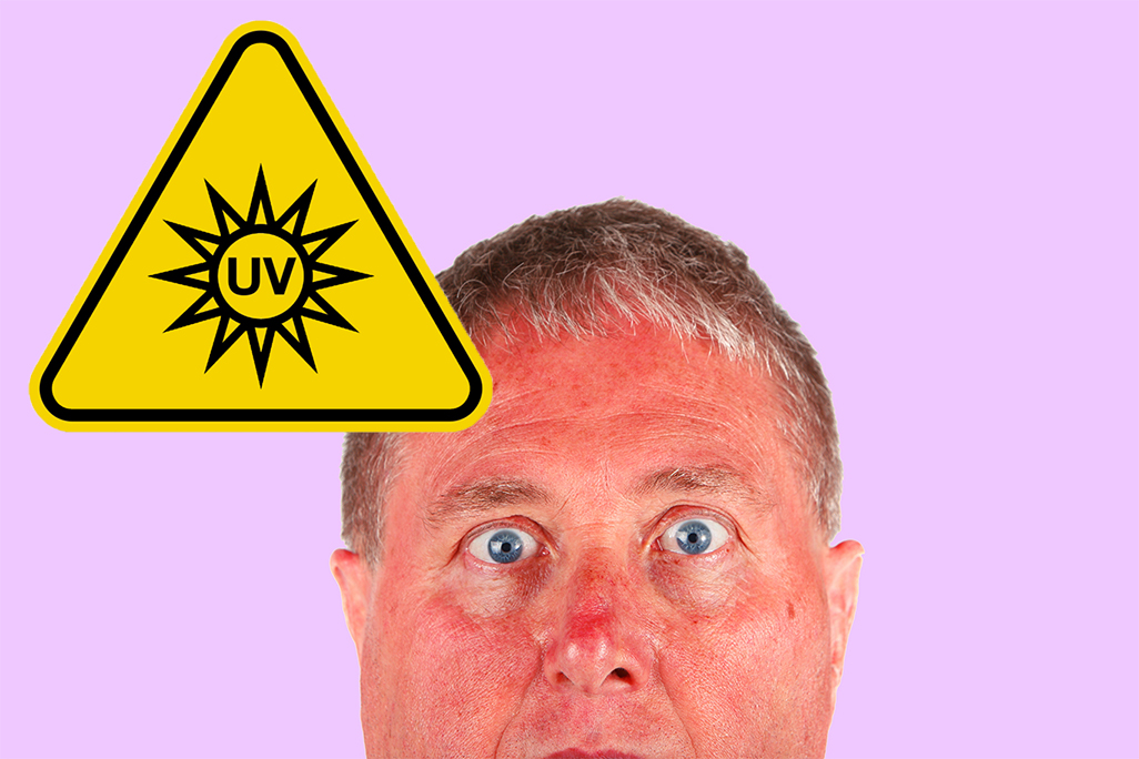
Dangerous for the user
Exposure to UV-light can cause serious damage to skin and eyes. During the time of usage, the viewers eyes and exposed skin should always be covered by protective wear.
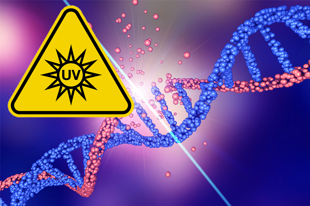
Dangerous for the sample
The quick destruction of sample DNA by UV-light can have severe effects on downstream applications. 30 s of UV-light expoure are enough to drastically reduce cloning efficiency!
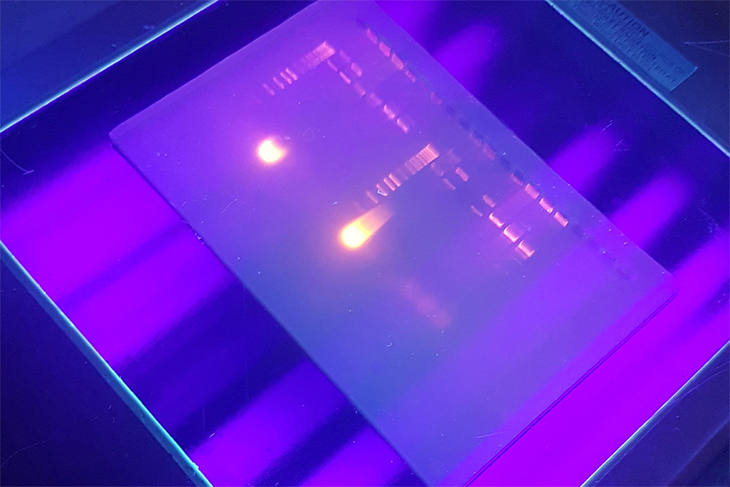
Lower efficiency
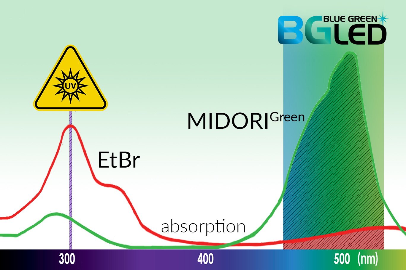
UV-light transilluminators only use a single excitation wavelength. This calls for lost excitation potential, compared to the spectral Blue/Green LED light (470 nm - 520 nm).
Blue/Green LEDs - Safe and powerful DNA detection
Blue/Green LEDs emit safe visual light at 470 nm - 520 nm. This light spectrum has no destructive properties for DNA, making the gel documentation process for the user as safe as watching TV, and keeping the DNA sample unharmed. Compared to UV-light, DNA integrity and cloning/sequencing efficiency is not at all affected, even after long light exposure times. The reassuring safety of Blue/Green LED light does not compromise its performance. Absorption spectra of most nucleic acid staining agents show sufficient light absorption in the blue/green light range. Due to the accumulated energy absorption of the entire blue/green spectral light, the powerful LED technology efficiently excites all commonly used red and green DNA dyes. Green DNA dyes, showing a very high absorption of blue/green light, show exceptional signals with superb band intensities. Our Blue/Green LED transilluminators show a remarkably stable and reliable light performance. With an LED life time of 10 years continuous illumination, or up to 20 years normal laboratory usage, the transilluminators represent robust, cost saving and sustainable systems.

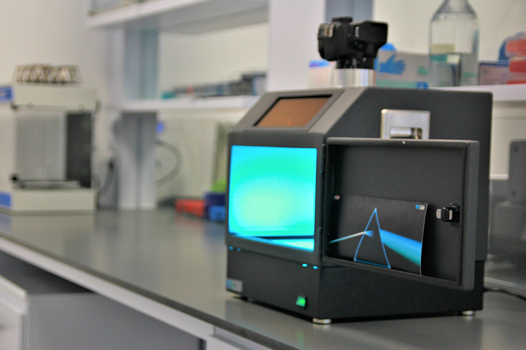
Safe use
Blue/Green LEDs are completely safe for everyday laboratory use. The LED light is emitted in the visible spectrum and does not cause any damage for the sample or for the user.
Efficient technology
What counts is the excitation area under the absoption curve! The Blue/Green spectral light efficiently excites red and green DNA dyes through accumulated energy absorption.

Cost saving
LED-lights are very stable and reliable and can be used for up to 20 years of normal laboratory handling. UV-light bulbs rapidly lose their efficiency and need to be replaced regularly.
Our Blue/Green LED Gel Documentation Systems
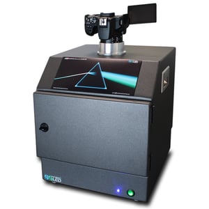
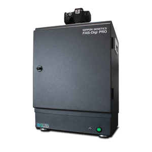
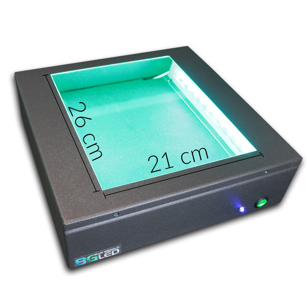
Our gel documentation systems are equipped the huge Blue/Green Transilluminator XL. Twelve powerful LEDs, positioned at each side, ensure a bright and evenly distributed illumination. The huge light area of 21x26 cm allows convenient gel positioning of any gel size. The Blue/Green Transilluminator XL is the core part of our FAS-DIGI Compact, FAS-DIGI PRO and FAS-V Gel Documentation Systems. It efficiently excites all common red and green DNA dyes with a very high intensity, making the systems highly impressive gel analysis tools.
FAS-DIGI Compact
- The compact stand-alone Gel Doc System is perfectly suited for limited lab space.
- It uses a high resolution scientific grade camera and is made from high quality coated aluminium.
- More information ...
FAS-DIGI PRO
- The advanced Gel Doc system includes a full control imaging software and a white LED plate for protein gel documentation.
- The attachable amber filter allows easy gel viewing and cutting.
- More information ...
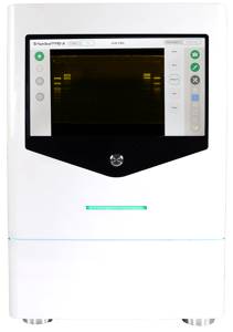
NEW | FAS-X
Gel imaging WITHOUT LIMITS
Unbeatable performance –With any fluorescent dye
